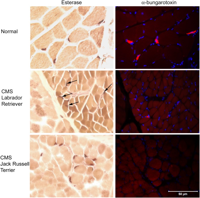
Figure 4. Cryosections (8 µm) of intercostal muscle from a normal dog, a Labrador Retriever with CMS (end-plate AChE deficiency), and a Jack Russell Terrier with CMS due to AChR deficiency (neuromuscular disease control) are illustrated.
For each dog, histochemical staining for esterase activity (brown stain) is shown along with a serial section demonstrating immunofluorescent localization of α-bungarotoxin for AChR and end-plate localization (red color). Muscle nuclei are blue (Dapi stain). There is a good correlation between esterase staining (brown) and α-bungarotoxin localization (red) in the control dog muscle. Although esterase staining is present in the Labrador Retriever muscle (arrows), the localization correlates poorly with that of AChRs. In the CMS Jack Russell Terrier esterase staining was present; however, staining for AChR was markedly decreased or absent, consistent with a markedly decreased AChR content. Bar = 50 µm for all images.

