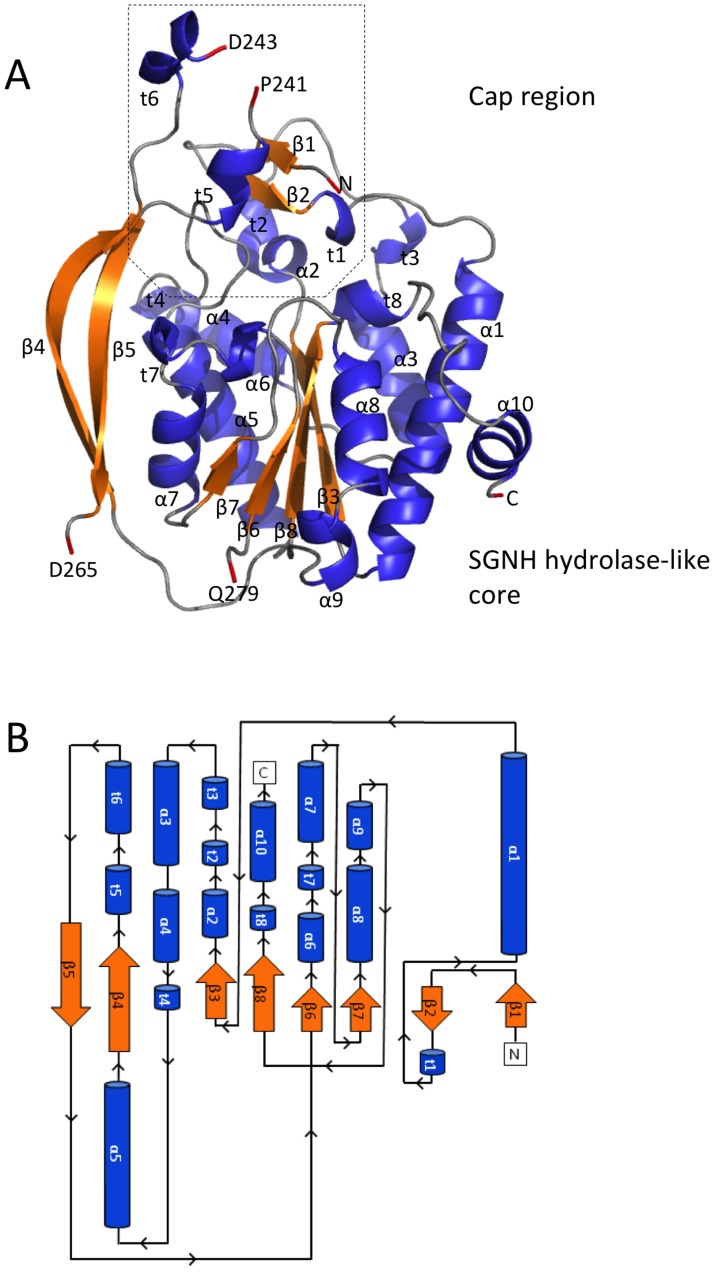
Figure 1. Structure and topology of PpAlgJ75–370.
(A) Cartoon representation of PpAlgJ75–370 with secondary structural elements labelled (α: α-helix; β: β-strand; and t: 310 helix). Residues at discontinuous points in the structure due to poor observed electron density are labelled and coloured red. The N- and C-termini of the protein are labelled N and C, respectively, and the terminal residue is coloured red. (B) Topology model of the PpAlgJ75–370 structure with secondary structural elements and termini labelled as in panel (A).

