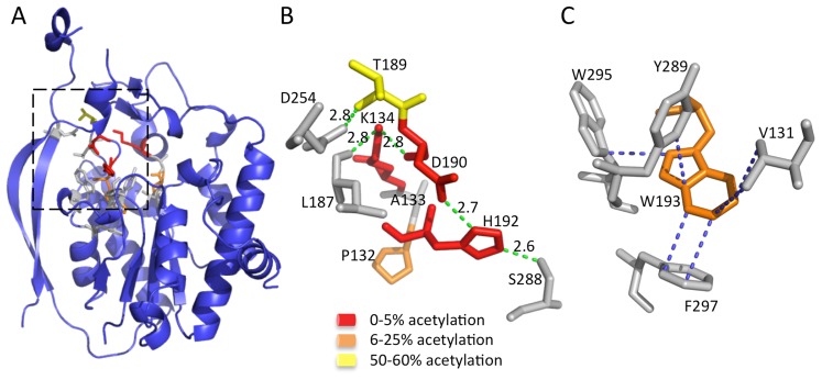
Figure 4. The AlgJ signature motifs.
(A) Cartoon representation of PpAlgJ75–370 with residues from the signature motif represented as sticks. The AlgJ signature motif residues have been coloured according to the amount of alginate acetylation observed in vivo in each alanine variant relative to WT AlgJ. Yellow represents impairment of between 50–60% acetylation; orange represents strong impairment with only 6–25% acetylation observed; and red represents ablation or between 0–5% observed acetylation. Residues represented as grey sticks are proposed to be involved in interactions with the AlgJ signature motif residues but have not been characterized in vivo. (B) Selected residues from the box area in panel A that participate in or are proposed to be involved in the hydrogen bonding network and are coloured as described in (A). Hydrogen-bonds are depicted as green dashes with distance given in angstroms (Å). (C) Residues participating in hydrophobic and van der Waals interactions with W193 (shown in orange) are depicted in grey. Hydrophobic and van der Waals interactions less than 3.7 Å are depicted with blue dashes.

