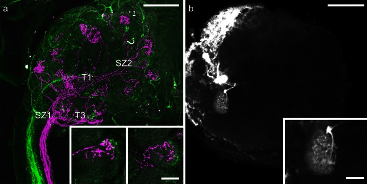
Fig. 2.
a Z-projection of a double-mass-staining of hair-like sensilla on segment 9 (magenta) and segment 4 (green) of honey bee antenna. The input tracts T1 and T3 are indicated. Note the two sorting zones (SZ1, SZ2). Bar 100 μm. Insets Detailed views of two glomeruli that are innervated by axons from olfactory receptor neurons from both the distal and the proximal parts of segment 9 in a layered fashion. Bar 25 μm. b Z-projection of an antennal lobe with an intracellularly stained projection neuron innervating a single glomerulus. Bar 100 μm. Inset Stained glomerulus in more detail; the dendritic arborizations of a projection neuron ramify throughout the entire glomerulus. Bar 25 μm

