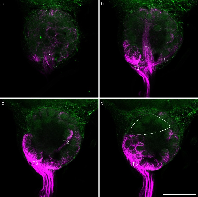
Fig. 3.
Images of a representative mass-staining of the axons of olfactory receptor neurons in Sensilla trichoidea of segment 5 of the honeybee worker antenna. Axons of olfactory receptor neurons are shown in magenta; the background was visualized via autofluorescence of the tissue and is shown in green. a Z-projection of 20 μm of the dorsal part of the stained antennal lobe; the glomeruli innervated from T1 input tract (T1) are clearly visible. b Z-projection of 20 μm of the dorsal middle part of the stained antennal lobe. Glomeruli innervated from the T3 input tract (T3) and the T1 input tract (T1) are clearly visible. c Z-projection of 20 μm of the ventral middle part of the stained antennal lobe. Glomeruli innervated from the T3 input tract (T3) and the T2 input tract (T2) are clearly visible. d Z-projection of 20 μm of the ventral part of the stained antennal lobe. Glomeruli innervated from the T3 input tract (T3) are clearly visible; the only non-innervated glomeruli (probably associated with T4 input tract) are indicated by the dashed circle. Bar 200 μm

