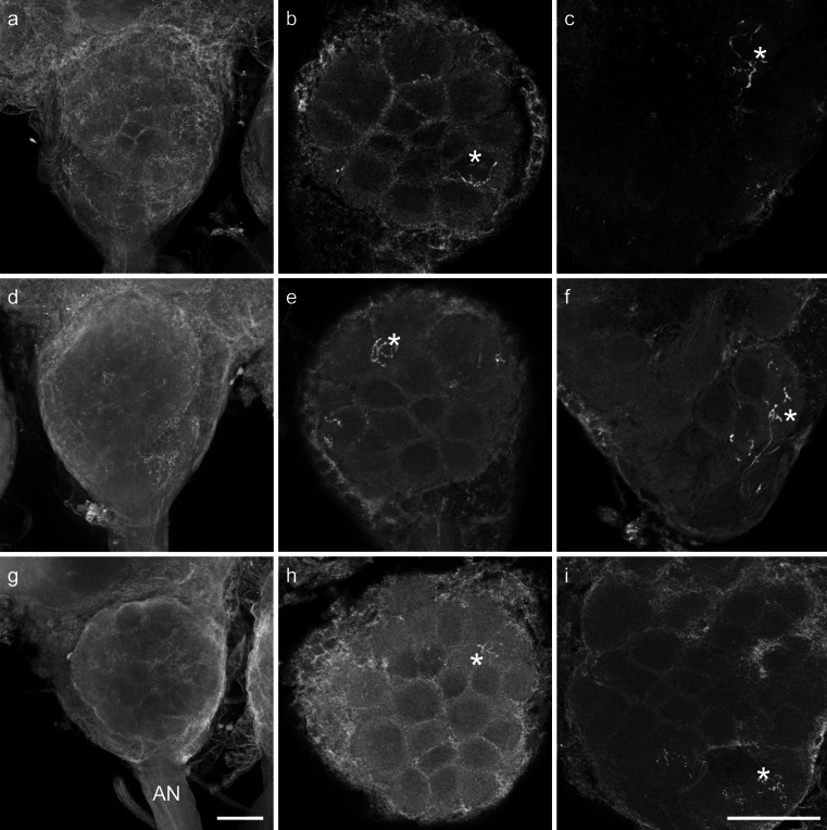
Fig. 4.
Confocal image stacks from two antennal lobes after staining of Sensilla basiconica-rich regions of the antenna (a–f) and image stacks of a single-sensillum staining of a S. basiconicum (g–i). Stained glomeruli (asterisks). a, d, g Complete stacks of antennal lobes with the antennal nerves (AN) and olfactory receptor neuron (ORN) arborizations in single glomeruli can hardly be identified in the complete image stacks. Bar 100 μm. b, e, h Substacks of the antennal lobes with identifiable ORN innervation in individual glomeruli in the T1 glomerular cluster. c, f, i Substacks of the antennal lobe with identifiable ORN innervation in individual glomeruli in the T3 glomerular cluster region. Bar 100 μm

