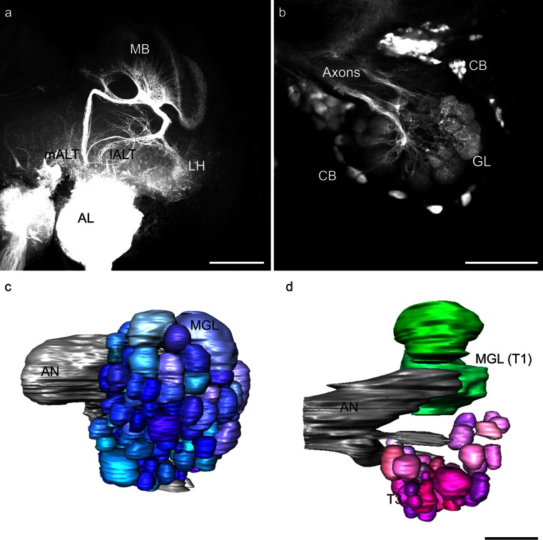
Fig. 6.
a Z-projection of a mass-staining of antennal lobe (AL) projection neurons in the honeybee drone; the AL, the medial and the lateral AL tract (mALT, lALT) and arborizations in the mushroom bodies (MB) and the lateral horn (LH) are visible. Bar 200 μm. b Z-projection of mALT staining in a honeybee drone. The axons, cell bodies (CB) and dendritic glomerular (GL) innervation are visible. Bar 100 μm. c Three-dimensional reconstruction of a drone AL with the antennal nerve (AN) after staining of all olfactory receptor neuron axons. Two macroglomeruli (MGL) are indicated. d Reconstruction of the mALT proportion of glomeruli and two macroglomeruli (MGL) within the T1 glomerular cluster (T1) as landmarks in a drone AL. The two lALT-associated MGL are shown in green, whereas mALT glomeruli in the T3 glomerular cluster (T3) are shown in shades of magenta. Bar 100 μm

