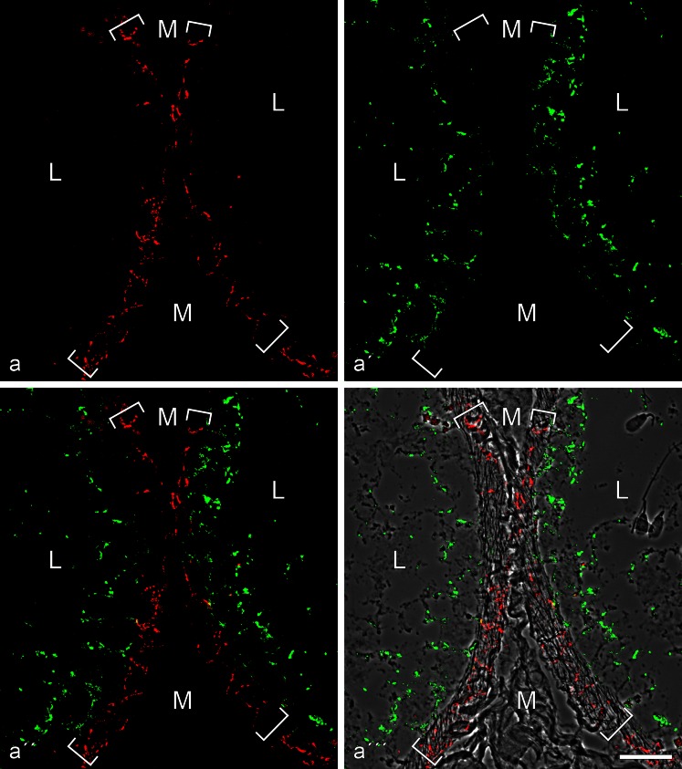
Fig. 4.
Double-label immunofluorescence microscopy of cryostat cross-sections through tubuli seminiferi of frozen bull testis after reactions with antibodies against E-cadherin (red; a, a'', a''') and N-cadherin (green; a', a'', a'''). In a''', the reactions are shown on a phase contrast background. Note the mutually exclusive immunostaining of cell-cell junctions of the adherens type, N-cadherin-based ones (green) in the Sertoli cells and spermatogonia of the tubuli seminiferi and E-cadherin-containing junctions exclusively in a special layer of myoid cells surrounding the tubuli (demarcated by the parentheses). M mesenchymal region with interstitial cells; L lumen of the seminiferous tubules, with individual spermatids (e.g., on the right-hand side of a'''). Bar 20 μm

