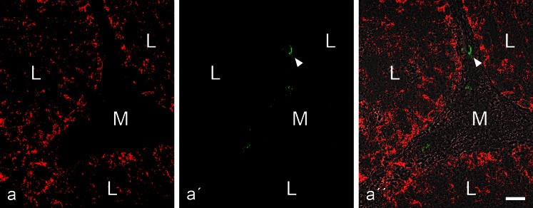
Fig. 5.
Double-label immunofluorescence microscopy showing the reactions of antibodies to N-cadherin (a, red, mouse mAb) and desmoplakin (b, green, guinea pig antibodies) on a cross-section through tubuli seminiferi of bull testis (L, tubular lumen; M, mesenchymal space). While the N-cadherin reaction identifies the AJs of the Sertoli and spermatogonial cell layer (a and a'' show the reaction in the three neighbouring tubular structures) there is no desmoplakin reaction (a'; for a visualization “control” this picture has been selected as, accidentally, a very small artifical green particle is seen here, denoted by a white arrowhead). Bar 20 μm

