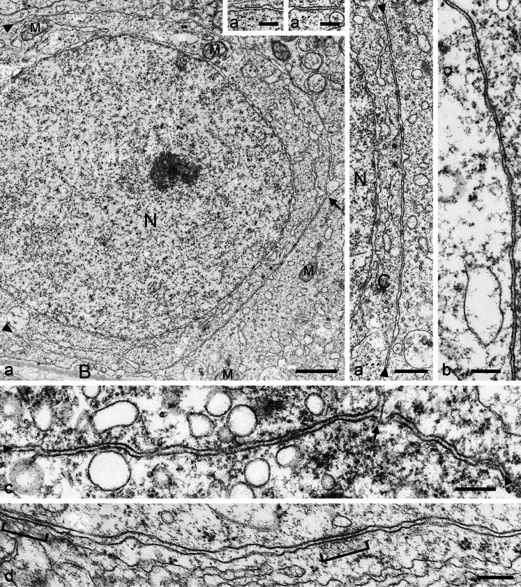
Fig. 9.
Electron micrographs of ultrathin sections through seminiferous tubules of boar (a–a''') and bull (b–d) testis, showing a survey picture including large parts of a nucleus (N) and details of the very extended, rather regularly narrow-spaced (membrane-to-membrane interspace 8–18 nm) area adhaerens junctions; B, basal lamina; M, mitochondrion (a'–a''' present details at higher magnification; C, cytoplasm). Such extended, narrow-spaced plasma membrane connections of the “minimal plaque material” AJ type are also seen in bovine Sertoli cells (b–d) and only occasionally rather thin, loosely and irregularly arranged plaque-like structures are detected (see, e.g., d, parentheses). Note that these MPM-AJ associations are also maintained at sites where the plasma membranes of three cells meet (c, arrow). Bars (a) 1 μm, (a''') 500 nm, (a', a'', b–d) 200 nm

