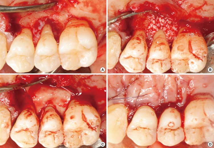
Figure 3.
Representative intraoperative photographs of guided tissue regeneration procedure using GuidOss (NIBEC, Jincheon, Korea). (A) A deep intrabony defect on the upper second premolar was detected. The defects and root surfaces were thoroughly planed and debrided. (B) The bone mineral was applied on the intrabony defect. (C) After grafting of bone mineral, a porous nonchemical cross-linking collagen membrane (GuidOss) was trimmed and adapted over the defect. The entire defect and 2-3 mm of the surrounding alveolar bone were completely covered with a membrane. (D) The flap was sutured and completely closed without membrane exposure.

