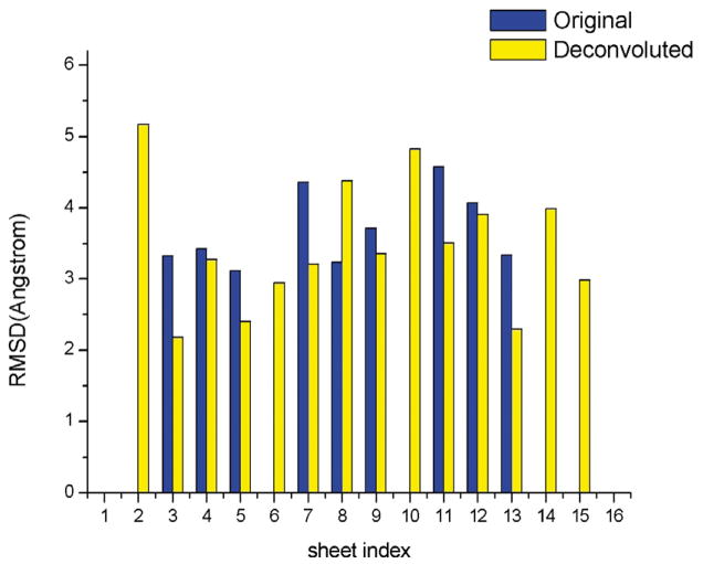
Figure 8.
Comparison of sheet-tracing results in the 7.6 Å density maps of the λ2 protein of reovirus with (yellow bar) and without (blue bar) deconvolution. There are a total of 16 β-sheets, 12 of which are large (three-stranded or more) and four are small (short two-stranded). In all but one (sheet 8) case, the deconvolution resulted in smaller rms deviations relative to the crystal structure than without. Moreover, the deconvolution brought up five additional β-sheets (sheets 2, 6, 10, 14, and 15) for which no pseudo-Cα traces could be built on the original maps without deconvolution.

