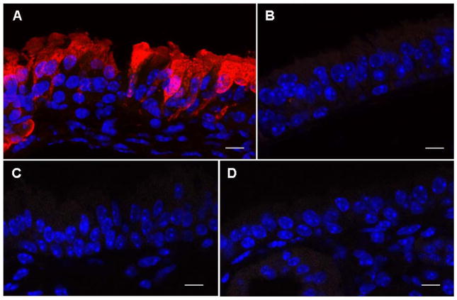
Figure 5. Detection of GFP-CFTR fusion protein in mouse nasal epithelium.
The nasal epithelium of CF knockout mice was perfused with pCIKGFPCFTR complexed to GL67A plus 0.5% CMC. A recombinant Sendai virus (SeV) carrying GFP-CFTR was used as positive control and untransfected animals were negative controls. 24 hr after transfection nasal tissue was processed for immunohistochemistry and expression of GFP-CFTR fusion protein was visualized using an anti-GFP antibody and a secondary antibody conjugated to AlexaFluor 594. (A) SeVGFP-CFTR with anti-GFP primary antibody, (B) SeV-GFP-CFTR without anti-GFP primary antibody, (C) pCIKGFP-CFTR with anti-GFP primary antibody, (D) untransfected mouse with anti-GFP primary antibody. GFP-CFTR protein appears in red. DAPI stained nuclei appear in blue. Representative images are shown. n=10 mice/group. Scale bar=10 μm.

