
Figure 2.
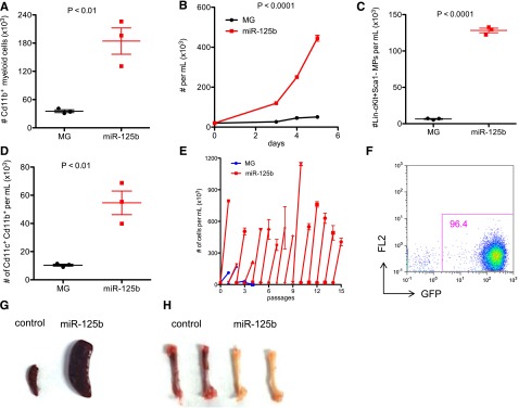
Overexpressing miR-125b in BMCs induces myeloproliferative disorder in vitro. (A) HSPC-enriched BMCs, which were obtained by injecting C57bl/6 mice with 5-fluorouracil, were transduced with MG control or MG-125b retroviruses. Equal numbers of BMCs were cultured in 50 ng/mL SCF, 50 ng/mL IL-6, and 25 ng/mL IL-3. After 5 days, the total number of myeloid cells (CD11b+) cells was determined by flow cytometry. (B) MG control or miR-125b–overexpressing HSPC-enriched cells were expanded at same starting density in 50 ng/mL SCF. Cells were counted by flow cytometry. (C) Equal numbers of HSPC-enriched cells transduced with MG or miR-125b overexpression cassette were expanded in 50 ng/mL SCF. The density of Lin−Sca1−cKit+ cells after 4 days was determined by flow cytometry. (D) Equal numbers of MG control or miR-125b–overexpressing BMCs were cultured in 20 ng/mL GMCSF to induce differentiation into dendritic cells. The number of dendritic cells was determined by flow cytometry 6 days after culture. (E) MG or miR-125b–transduced BMCs were cultured in 50 ng/mL SCF. When the miR-125b–overexpressing cells were confluent between 4 and 8 days after plating, the cells were reseeded at a starting density of 20K per mL. The x-axis represents the passage number, and the y-axis represents the cell density. Panels A through E are representative of at least 2 independent experiments. (F) MiR-125b–overexpressing BMCs were cultured and expanded in vitro. One million GFP+ cells were injected into sublethally irradiated C57bl/6 mice. Two months posttransplantation, recipient blood was subjected to flow cytometric analysis to determine engraftment of GFP+ cells. The pictures of (G) spleen and (H) femur are representative examples of these organs taken from moribund miR-125b–transduced mice (right panel) and age-matched C57bl/6 controls (left panel). Representative of 6 mice.