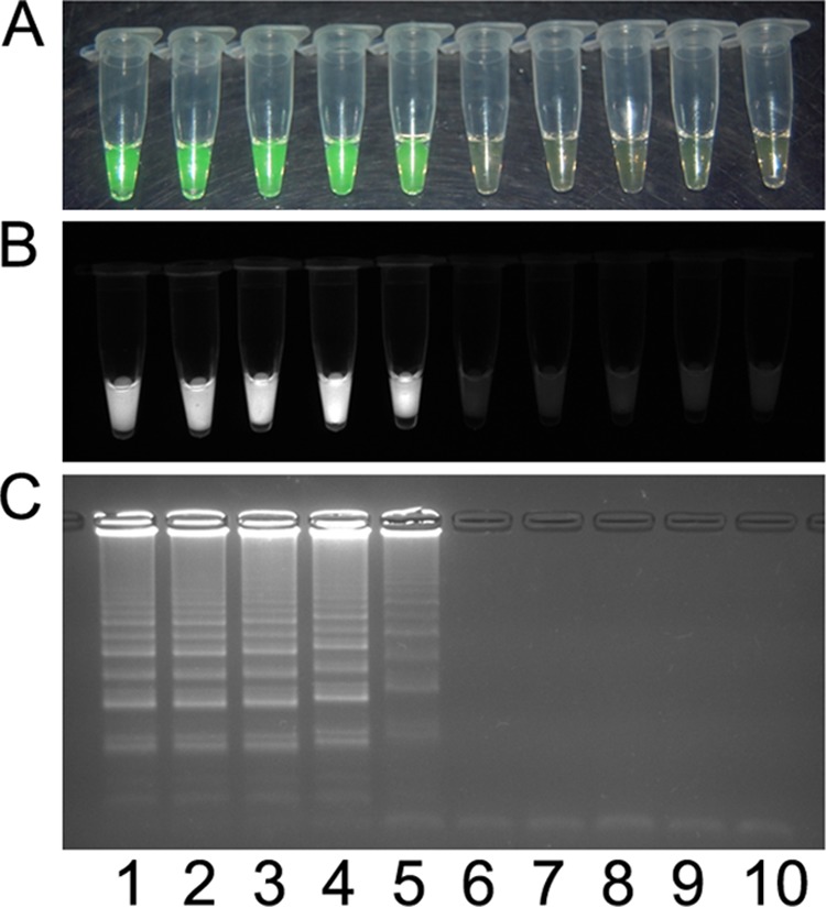
FIG 5.

Visual detection of X. oryzae pv. oryzae PXO99A heat-killed cells using the pathovar-specific LAMP assay. The assay was used to test a dilution series consisting of 108 CFU ml−1 (lane 1), 107 CFU ml−1 (lane 2), 106 CFU ml−1 (lane 3), 105 CFU ml−1 (lane 4), 104 CFU ml−1 (lane 5), 103 CFU ml−1 (lane 6), 102 CFU ml−1 (lane 7), and 101 CFU ml−1 (lane 8); X. oryzae pv. oryzicola BLS256 (lane 9); and a no-template control (lane 10). Products were detected by using 1 μl Quant-IT Pico green reagent (Life Technologies, Grand Island, NY) under visual light, where a positive result changes from orange to green (A), or ultraviolet light, where a positive result fluoresces (B), or by 1.5% agarose gel electrophoresis, where a positive result is a laddered product (C).
