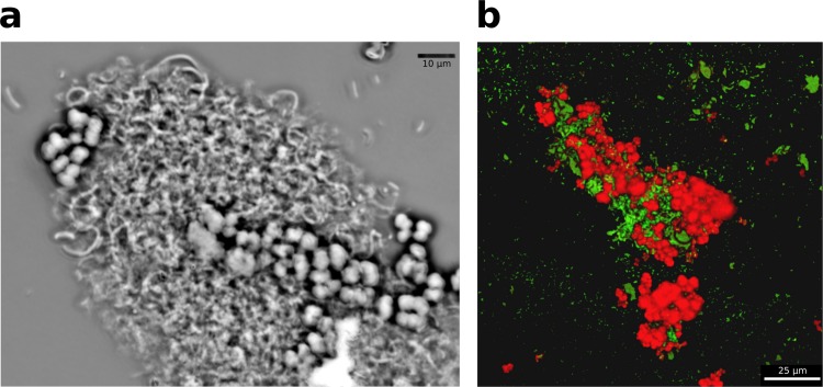
FIG 2.
Coculture composition. (a) Phase-contrast micrograph of an aggregate of M. barkeri (cocci) and G. metallireducens (rods). (b) Scanning laser confocal microscope image of coculture aggregate after in situ hybridization, which targeted Methanosarcina cells with a red probe (Cy3) and G. metallireducens cells with a green probe (Cy5). Images are representative of triplicate samples taken during the mid-exponential growth phase of the cocultures.

