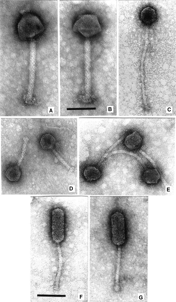
FIG 2.
Electron micrographs. (A) Listeria phage LP-083-2 with an extended tail and hexagonal head. The presence of capsomers is hinted at by small asperities on the capsid. (B) LP-083-2 with a pentagonal head. (C) Phage LP-030-3. (D) Two particles of phage LP-083-1. (E) Three particles of phage LP-030-2. (F) Listeria phage LP-032. (G) Enterococcus phage VD13. Phages were stained with uranyl acetate and visualized at a final magnification of ×297,000. Panels A to E and panels F and G, respectively, are shown at the same scale; bars indicate 100 nm.

