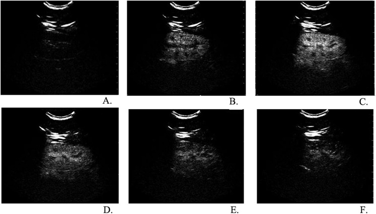
Figure 1.
Real-time observation of renal cortical perfusion in different stages after a bolus injection of SonoVue® (Bracco Imaging S.p.A., Milan, Italy); from the longitudinal plane, we observed a quick and intense enhancement of the cortex (a). Initial visualization of the contrast agent occurred after 15–25 s, from segmental renal arteries to small interlobular arteries and immediately followed by an intense and uniform enhancement of the renal cortex (b). After the enhancement of the cortex, the pyramids were gradually filled in with contrast agents and became isoechoic with the cortex. In about 30–45 s, enhancement of the renal cortex reached peak intensity (c). In 50–180 s, the renal cortex enhancement gradually decreased as the contrast concentration decreased (d–f). No significant delay was observed in the perfusion of the renal cortex between patients with early chronic kidney dysfunction and the control group.

