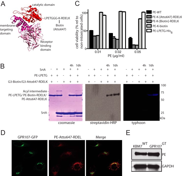
FIGURE 2.
PE is transported through the Golgi to reach the ER. A, crystal structure of PE depicting its three domains and the C-terminal site where biotin or fluorophore is conjugated using sortase-mediated labeling strategy. B, sortase-catalyzed attachment of biotin or Atto647 dye to PE. Reactions were analyzed by SDS-PAGE with visualization by Coomassie gel, streptavidin-HRP blot, and typhoon image showing PE was successfully labeled with biotin or Atto647 bearing RDEL at the C terminus. See “Experimental Procedures” for details. C, HeLa cells were intoxicated with different concentrations of PE-WT, PE-without RDEL motif (PE-LPETG or PE-LPETG-K-biotin) or labeled PE carrying RDEL motif at the C-terminal (PE-(biotin)-RDEL or PE-(Atto647)-RDEL), and cell viability was determined and shown as percent relative to nonintoxicated cells. Error bars represent S.D. of three experiments performed in duplicate. D, confocal images of HeLa cells expressing C-terminally GFP-tagged GPR107 that were intoxicated with PE-(Atto647)-RDEL. Note that PE partially colocalizes with GPR107. E, similar number (1 × 10⁶) of GPR107 null and wild type KBM7 cells were intoxicated with 50 ng/ml PE for 30 min, and the cell lysates (∼20 μg) were analyzed by immunoblots using PE and GAPDH antibodies.

