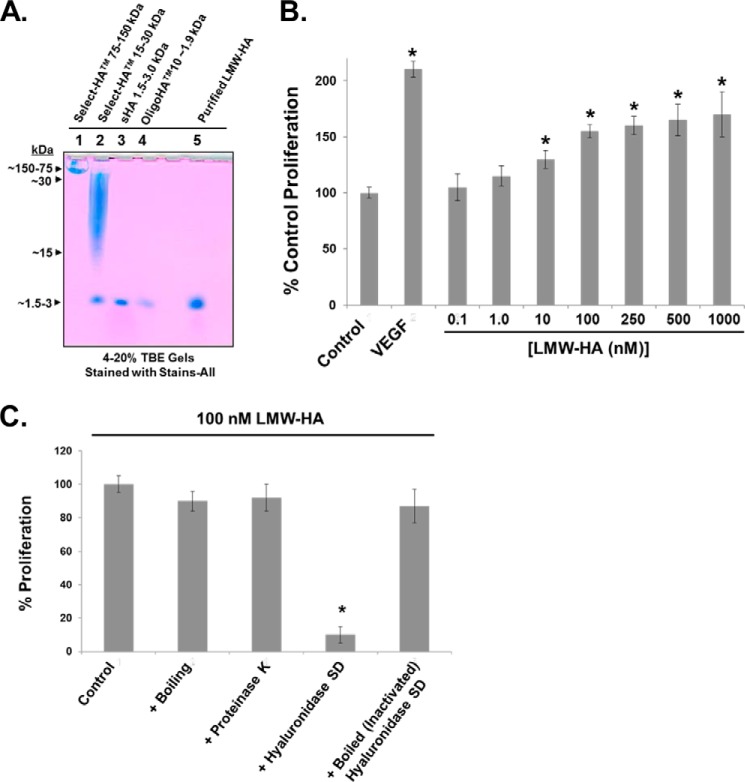
FIGURE 1.
Characterization of LMW-HA and its effects on EC proliferation. Panel A, to evaluate the molecular weight (MW) and purity of our LMW-HA preparation, a 4–20% Tris borate-EDTA (TBE) gel was run with HA molecular weight standards (Select-HATM 75–150 kDa (Sigma), Select-HATM 15–30 kDa (Sigma), small HA 1.5–3.0 kDa (Enzo Life Sciences), and OligoHATM10 ∼1.9 kDa (Hyalose, L.L.C.)) and our purified LMW-HA (see “Experimental Procedures”). Development of the resulting gel was achieved with Stains-All (Sigma). Panel B, determination of the minimal molar concentration of our purified LMW-HA to induce maximal HLMVEC proliferation. Control, VEGF (200 pg/ml), or LMW-HA (0.1–1000 nm) pretreated EC (5 × 10³ cells/well) were incubated with 0.2 ml of serum-free media for 72 h at 37 °C in 5%CO2, 95% air in 96-well culture plates. The in vitro cell proliferation assay was analyzed by measuring increases in cell number using the CellTiter96TM MTS assay (Promega) and read at 492 nm. Each assay was set up in triplicate and repeated at least five times. An asterisk indicates a statistically significant difference (p < 0.05) from Control. Panel C, to further test the purity of our purified LMW-HA, it was subjected to boiling and proteinase K digestion (50 μg/ml), which did not affect the ability to promote EC proliferation compared with untreated 100 nm LMW-HA. LMW-HA was also fully digested with hyaluronidase SD (100 milliunits/ml), and there was virtually no increase in EC proliferation, a process that was reversed with boiling (inactivating) hyaluronidase SD before HA digestion. Each assay was set up in triplicate and repeated at least five times.

