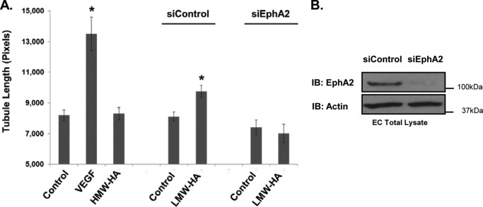
FIGURE 6.
Silencing of EphA2 blocks LMW-HA-induced angiogenesis. Panel A, EphA2 and control silenced cells were seeded on Matrigel-coated plates in the presence or absence of LMW-HA (100 nm). Images were captured after a 6-h incubation, and tubule formation was quantified using ImageJ image analysis software. The mean tubule length was calculated for 20 images per treatment and graphically depicted. The asterisks (*) indicate a statistically significant difference (p < 0.05) from control. No significant difference was detected between siEphA2 and siEphA2 LMW-HA. Each treatment was performed in triplicate, and experiments were repeated three times. Results are expressed as tubule length per treatment. Panel B, immunoblot (IB) analysis of HLMVEC treated with EphA2 siRNA or control siRNA. Cell lysates were blotted using EphA2 and actin antibodies to confirm EphA2 silencing.

