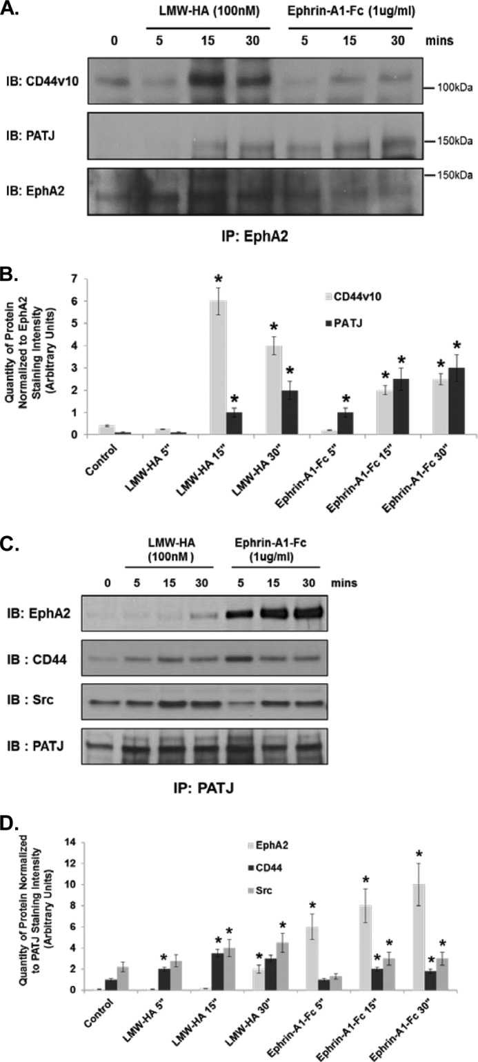
FIGURE 7.

LMW-HA stimulation of HMVEC induces CD44, EphA2, PATJ, and Src complex formation. Panel A, ECs were serum-starved for 1 h in EBM-2 media and then incubated with LMW-HA (100 nm) or Ephrin-A1/Fc (10 ng/ml) for 5, 15, and 30 min. Cell lysates were prepared in 1% Nonidet P-40 lysis buffer and incubated with anti-EphA2-cross-linked beads for immunoprecipitation. The resulting immune precipitations (IP) were then blotted for CD44v10, PATJ, and EphA2. IB, immunoblot. Panel B, graphical depiction of quantity of protein normalized to EphA2 staining intensity (arbitrary units) from experiments described in panel A performed in triplicate and quantitated using computer-assisted densitometry. The asterisks (*) indicate a statistically significant difference (p < 0.05) from control. Panel C, ECs were serum-starved for 1 h in EBM-2 media and then incubated with LMW-HA (100 nm) or Ephrin-A1/Fc (10 ng/ml) for 5, 15, and 30 min. Cell lysates were prepared in 1% Nonidet P-40 lysis buffer and incubated with anti-PATJ-cross-linked beads for immunoprecipitation. The resulting immune precipitations were then blotted with EphA2, CD44, Src, and PATJ antibodies. Panel D, graphical depiction of quantity of protein normalized to PATJ staining intensity (arbitrary units) from experiments described in panel A performed in triplicate and quantitated using computer-assisted densitometry. The asterisks indicate a statistically significant difference (p < 0.05) from control.
