FIGURE 9.
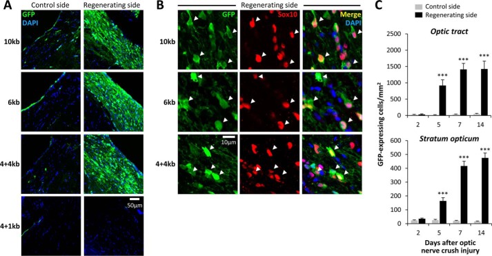
The mpz AIRE is dissociable from the mpz upstream oligodendroglial enhancer. A, expression of GFP in the ventral optic tract of Tg(mpz:egfp) zebrafish. Each row of confocal micrographs shows a different transgenic line expressing GFP under transcriptional control of distinct fragments of the mpz regulatory region (indicated to the left; see Fig. 8A). The columns show control optic tract (left) and regenerating optic tract (right) at 14 days postinjury. All panels are labeled for GFP (green) and DAPI (blue). The scale bar for all eight panels is shown in the bottom right panel. B, single confocal planes through the regenerating optic tract are shown at high magnification for the same transgenic lines as shown in A. The scale bar for all nine panels is shown at the bottom left. Sections are labeled for GFP (green; first column) and Sox10 (red; second column). The third column shows the merged image with a nuclear counterstain (DAPI, blue). The arrowheads show the positions of GFP-expressing cells, confirming that they were also immunoreactive for Sox10. C, GFP-expressing cells in Tg(mpz[6kb]:egfp) zebrafish were quantified in the optic tract and stratum opticum at time points between 2 and 14 days after optic nerve crush injury, on each side of the same sections. The graphs show mean and S.E. (error bars). ***, p < 0.0001, control versus regenerating side; paired t test with Bonferroni correction for multiple comparisons.

