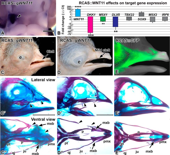
FIGURE 1.
Overexpression of WNT11 virus causes clefts. A, RCAS::WNT11 injected into the maxillary prominence at stage 16 and fixed 48 h later at stage 24. Expression of POL probe confirms the virus spread on the right side (arrowhead). B, quantitative PCR measurement of gene expression change in WNT11-infected maxillary tissue, 48 h post-infection. Fold differences in expression were calculated relative to the GFP controls. Asterisks indicate genes that were significantly different. (****, p < 0.00001; ***, p < 0.001; **, p < 0.01) than GFP control RNA. C–D″, stage 38 embryos with RCAS::WNT11 injected into the maxillary prominence at stage 15, photographed externally and after staining the skeleton. C–C″, an example of a severe phenotype with a notched upper beak or a cleft lip. The premaxilla is truncated and the palatine bone is shortened (arrowheads). The maxillary bone is displaced proximally (arrow). D–D″, an example of a mild phenotype with absent jugal bone (arrowheads). E–E″, a control embryo injected with RCAS::GFP at stage 15. The GFP signal is visible on the lateral side of the beak. The skeleton has developed normally. The abbreviations used are as follows: e, eye; ju, jugal bone; mx, maxillary prominence; mxb, maxillary bone; np, nasal pit; p, palatine; qj, quadratojugal; pmx, premaxilla; pt, pterygoid. Scale bars, A, 500 μm; Bar in C′ applies to C–F″, 5 mm.

