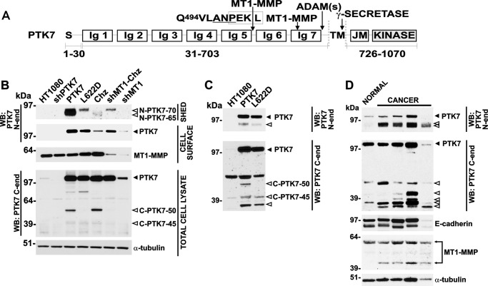
FIGURE 1.
PTK7 expression in cultured cells, tumor xenografts in mice, and colorectal cancer clinical samples. A, structure of the full-length PTK7 and the Chz mutant. MT1-MMP, ADAM, and γ-secretase cleavage sites are shown by arrows. The original MT1-MMP cleavage site (Pro-Lys-Pro↓Leu622) is localized in the seventh Ig-like domain of PTK7. The Chz mutation (Ala-Asn-Pro, underlined) results in an additional MT1-MMP cleavage site (Pro-Glu-Lys↓Leu512; boxed) in the junction region between the fifth and the sixth Ig-like domains of PTK7. S, signal peptide; Ig, immunoglobulin-like domain; TM, transmembrane domain; JM, juxtamembrane region; KINASE, the inert kinase domain. Residue numbering is shown below the structure. B, shed, soluble (Shed), biotinylated cell surface (Cell surface), and total cell lysate (Total Cell lysate) samples were analyzed using Western blotting with PTK7, MT1-MMP, and α-tubulin (loading control) antibodies. C, the status of PTK7 in tumor xenografts. Protein lysates were prepared from HT1080, PTK7, and L622D tumor xenografts developed in immunodeficient nu/nu mice. The lysate aliquots (30 μg/lane total protein each) were then analyzed using Western blotting with PTK7 antibodies directed to the N-terminal ectodomain (N-end) and to the C-terminal cytoplasmic tail (C-end). D, the status of PTK7 and MT1-MMP in human colon cancer tissue specimens. Tissue lysates (Protein Biotechnologies) prepared from colorectal tumors, and matching normal colonic tissues were analyzed by Western blotting with the PTK7, MT1-MMP, E-cadherin, and α-tubulin (loading control) antibodies. The two PTK7 antibodies we used were directed to the N-terminal ectodomain (N-end) and the C-terminal cytoplasmic tail (C-end). Black and open arrowheads indicate the full-length PTK7 protein and its proteolytic fragments, respectively. The enzyme and the C-terminal self-proteolytic fragment of membrane-tethered MT1-MMP are shown by the connected arrows. WB, Western blotting.

