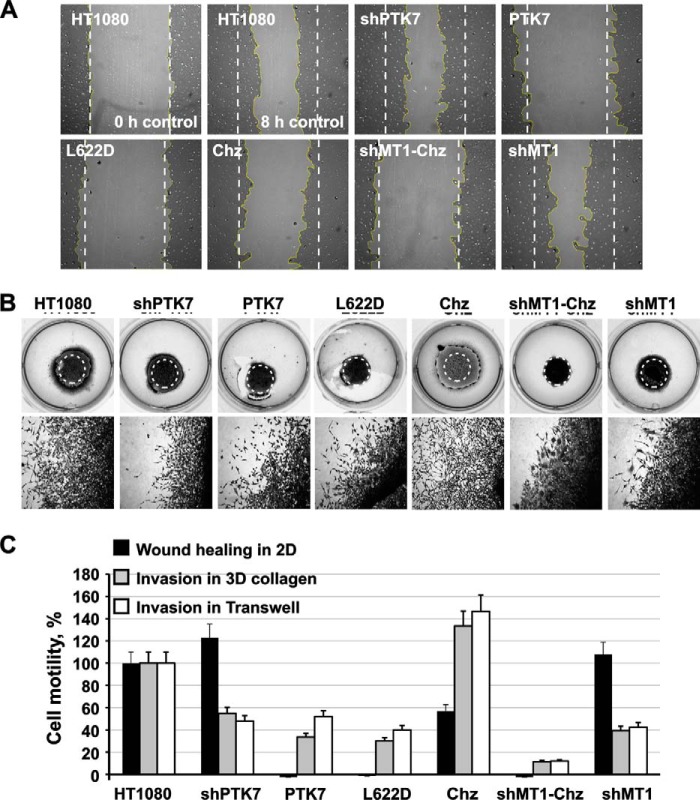
FIGURE 2.
PTK7 in cell locomotion. A, wound healing scratch assay. Following scratching of an artificial gap in a confluent cell monolayer, cells were allowed to migrate for 8 h to close the gap. The borders of the original scratch (time = 0) are shown by dotted lines. The yellow lines highlight cell borders after 8 h. B, cell invasion in three-dimensional collagen. Cells were embedded in collagen spheres in serum-free medium. Collagen solution that contained 10% FBS as a chemoattractant was then overlaid on the spheres and then also allowed to form a gel layer. Cells were allowed to migrate from the spheres and invade the collagen layer for 72 h. The original spheres (time = 0) are encircled. Cell images are shown at the bottom. C, relative locomotion of HT1080 (= 100%), PTK7, Chz, shPTK7, shMT1, and shMT1-Chz cells in the wound healing scratch assay and the invasion and invasion assays in the three-dimensional (3D) collagen matrix and transwells. The assays were performed in triplicate. ± S.D. (error bars) did not exceed 10%; p < 0.05.

