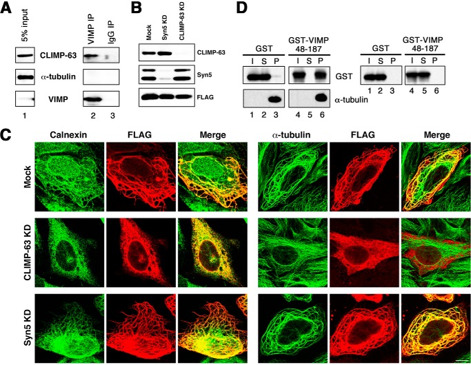
FIGURE 2.
VIMP interacts with both CLIMP-63 and polymerized MTs. A, membrane fractions of 293T cell lysates were prepared, solubilized, and immunoprecipitated (IP) with an anti-VIMP antibody (lane 2) or a control IgG (lane 3). The precipitates were separated by SDS-PAGE and analyzed by immunoblotting with antibodies against CLIMP-63, α-tubulin, and VIMP. 5% of solubilized membranes was run as input (lane 1). B and C, HeLa cells were transfected without (Mock) or with siRNA targeting Syn5 or CLIMP-63. At 48 h after transfection, the cells were transfected with the plasmid for FLAG-VIMP(1–187). After 24 h, the cells were lysed, separated by SDS-PAGE, and analyzed by immunoblotting with the indicated antibodies (B). Alternatively, the cells were fixed and double-stained with antibodies against FLAG and calnexin (C, left) or α-tubulin (right). KD, knockdown. Scale bar = 10 μm. D, purified GST (lanes 1–3) and GST-VIMP(48–187) (lanes 4–6) were subjected to sedimentation with (left) or without (right) Taxol-stabilized MTs and analyzed by immunoblotting with the indicated antibodies. I, S, and P represent input, supernatant, and precipitate, respectively.

