FIGURE 3.
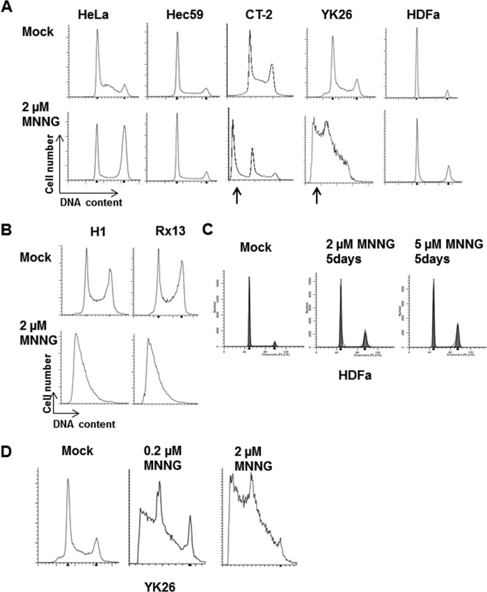
DNA alkylation damage induces apoptosis in human pluripotent stem cells. A, representative cell cycle profiles of HeLa, Hec59, CT-2, YK26, and HDFa cell lines with or without 2 μm MNNG for 48 h as measured by flow cytometry. The arrows indicate the presence of sub-G1 populations associated with apoptotic cells. B, representative cell cycle profiles of H1 and Rx13 cells with or without 2 μm MNNG for 48 h. C, representative cell cycle profiles of HDFa cells with or without 2 or 5 μm MNNG for 5 days. D, representative cell cycle profiles of YK26 cells mock-treated or treated with 2 or 0.2 μm MNNG.

