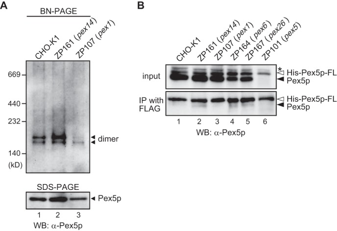
FIGURE 9.

In vivo oligomerization of Pex5p is impaired in pex1 ZP107 cells. A, cytosolic fractions each from CHO-K1 (lane 1), pex14 ZP161 (lane 2), and pex1 ZP107 (lane 3) were separated by blue native PAGE. Protein complexes were detected by immunoblotting with anti-Pex5p antibody. Molecular mass markers are on the left. BN, blue native; WB, Western blot. B, purified recombinant His-Pex5p-FLAG was introduced into CHO-K1 (lane 1), pex14 ZP161 (lane 2), pex1 ZP107 (lane 3), pex6 ZP164 (lane 4), pex26 ZP167 (lane 5), and pex5 ZP101 (lane 6) cells. Cells were then cultured for 24 h at 37 °C. His-Pex5p-FLAG was immunoprecipitated from cell lysates using anti-FLAG antibody. Immunoprecipitates were analyzed by SDS-PAGE and immunoblotting with anti-Pex5p antibody. Input, 10% input. Solid and open arrowheads indicate endogenous Pex5p and His-Pex5p-FLAG, respectively. *, a nonspecific band.
