
FIGURE 4.
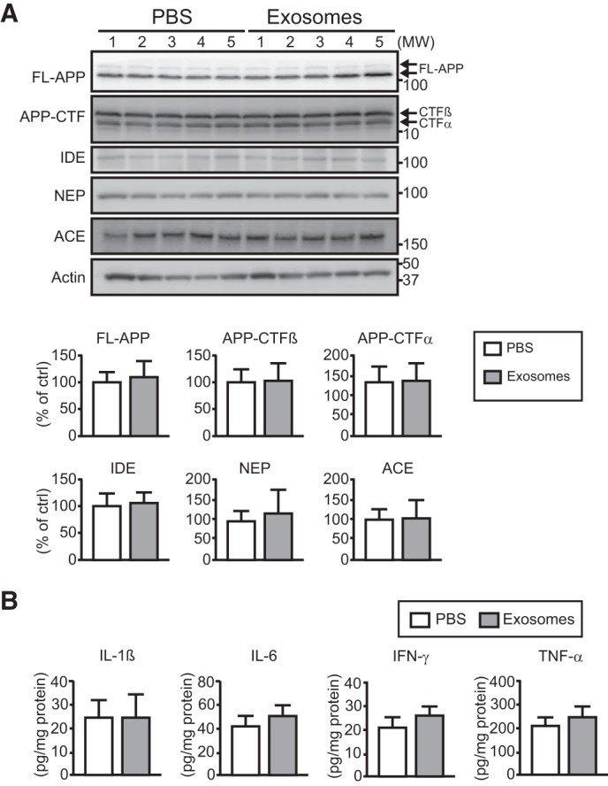
Exogenous exosomes do not stimulate APP processing, expressions of Aβ-degrading enzymes, or inflammatory response. Exosomes or PBS were continuously infused into the lateral ventricles of APP mice (4 months old) for 14 days (A and B). n ≥ 5 animals per group. A, full-length (FL)-APP, APP-CTFs, and the Aβ-degrading enzymes insulin-degrading enzyme (IDE), neprilysin (NEP), and angiotensin-converting enzyme (ACE) were detected in the hippocampus by Western blotting, and the intensity for each band was quantified. Data presented are the mean ± S.D. B, expression levels of proinflammatory cytokines (IL-1β, IL-6, IFN-γ, and TNF-α) in the hippocampus were measured by ELISA. Data are the mean ± S.D.