
FIGURE 6.
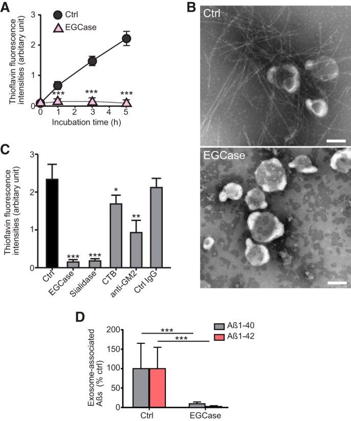
Exosomal GSLs are involved in Aβ assembly. A, thioflavin fluorescence intensities were measured in mixtures of exosomes (untreated as Ctrl or treated with EGCase) incubated with 15 μm Aβ1–42. Data are presented as the mean ± S.D.; ***, p < 0.001 (n = 4). B, representative electron microscopic images of exosomes incubated for 5 h with 15 μm Aβ1–42 are shown. Scale bar, 100 nm. C, the exosomes (untreated as Ctrl or treated with EGCase or sialidase) were incubated for 5 h with 15 μm Aβ1–42. The untreated exosomes were reacted with Aβ in the presence of cholera toxin B subunit (CTB) or anti-GM2 antibody. Fluorescence intensities of thioflavin-T were then measured. Values in each column are the mean ± S.D. of five values. *, p < 0.05; **, p < 0.01; ***, p < 0.001. D, biotinylated exosomes (untreated as Ctrl or treated with EGCase) stereotaxically injected into the hippocampus of APP mice (4 months) were isolated at 3 h after the injection, and the levels of exosome-associated Aβ were quantified by ELISA. Values are the mean ± S.D., ***, p < 0.001 (n = 4).