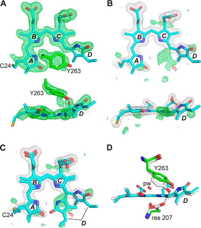
FIGURE 3.

Crystal structure of the DrBphP PAS-GAF fragment as Pr and bearing the D207A mutation. A, structure of BV at 1.75 Å resolution (PDB code 4Q0I) displayed in front and side orientations superimposed on the Fo − Fc electron density map generated by omitting BV and Tyr-263 and contouring to 3 σ. The final refined position of Tyr-263 is also shown. The sulfur atom in Cys-24 (yellow) that participates in the thioether linkage to BV is included, and pyrrole rings A–D are labeled. Carbons, nitrogens, and oxygens are colored cyan, blue, and red, respectively. B, BV is shown as in A, superposed with the final 2Fo − Fc (white) and Fo − Fc (positive, green; negative, red) electron density maps that were contoured at 1 σ and 3 σ, respectively. Atoms of the A and D pyrroles were refined at 70% occupancy, and atoms of the B and C pyrroles were refined at 100% occupancy. C, refined positions of alternate conformations of BV having the ZZZssa and ZZZsss configurations were superposed on the electron density maps from B. D, positions of Tyr-263 for wild-type PAS-GAF fragment and the D207A mutant were shown after superposition of the GAF domains. ZZZssa BV configuration, pyrrole water (pw), and residue (res) 207 positions are shown. Coloring scheme is the same as in A–C, except that wild-type carbons are white. Dashed line locates a hydrogen bond contact.
