FIGURE 5.

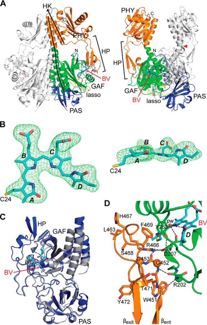
Crystal structure of the PSM dimer from DrBphP as Pr at 2.75 Å resolution. A, ribbon diagram of the PSM dimer (PDB code 4Q0J) in front and side views. For one subunit, the PAS, GAF, and PHY domains and the knot lasso are colored in blue, green, orange, and yellow, respectively. BV is located by the arrow and colored in cyan with the nitrogens and oxygens colored in blue and red, respectively. The hairpin (HP) extending from the PHY domain is located by the bracket. The lines track the helical spine of one monomer. Ala-326 at the apogee of the bowed helical spine is marked by a red arrowhead. HK, histidine kinase domain; N, N terminus; C, C terminus. B, ZZZssa conformation of BV displayed in front and side orientations superposed on the Fo − Fc electron density map generated by omitting BV and contouring to 3 σ. The sulfur atom in Cys-24 (yellow) that participates in the thioether linkage to BV is included, and pyrrole rings A–D are labeled. Carbons, nitrogens, and oxygens are colored cyan, blue, and red, respectively. C, superposition of the high resolution PAS-GAF structure (PDB code 4Q0H, gray) onto the PSM structure (blue). D, close-up view of the hairpin region (orange) extending toward and contacting the GAF domain close to the chromophore. The side chains of key amino acids are included, some of which are analyzed in Figs. 9 and 10. Dashed lines locate hydrogen bond contacts. The PSM structure is colored as in A. βent and βexit label the entrance (N-terminal) and exit (C-terminal) β-strands in the hairpin.
