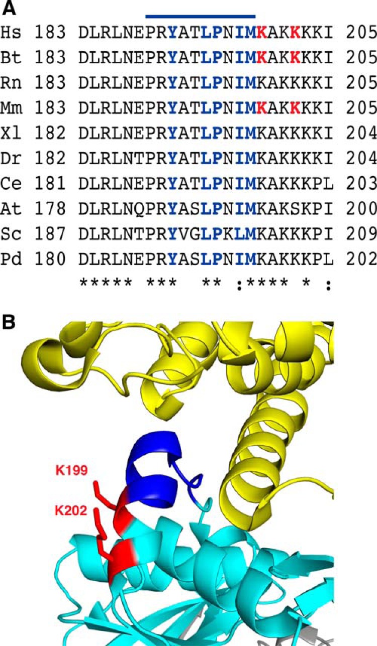
FIGURE 7.

Proximity of the methylated lysines to the recognition loop in ETFβ. A, conservation of sequences of recognition loops and adjacent residues from representative species. Hs, Homo sapiens; Bt, Bos taurus; Rn, Rattus norvegicus; Mm, Mus musculus; Xl, Xenopus laevis; Dr, Danio rerio; Ce, Caenorhabditis elegans; At, Arabidopsis thaliana; Sc, S. cerevisiae; Pd, Paracoccus denitrificans. The symbols (* and :) denote identical and conserved residues, respectively. The line above the sequence indicates the position of the recognition loop (33). Residues that interact with dehydrogenases are blue, and trimethylated lysines in the human, bovine, and mouse proteins are red. B, structure of the region of interaction between recombinant human ETFβ (turquoise blue) and the N-terminal region (extended region from residues 10–16 followed by an α-helix from residues 17–46) of the human medium-chain acyl-CoA dehydrogenase (yellow). The recognition loop (dark blue) consists of a loop (Pro189-Thr193) and part of an α-helix (Leu194-Met198) and is followed immediately by methylated lysines 199 and 202 (red).
