
FIGURE 5.
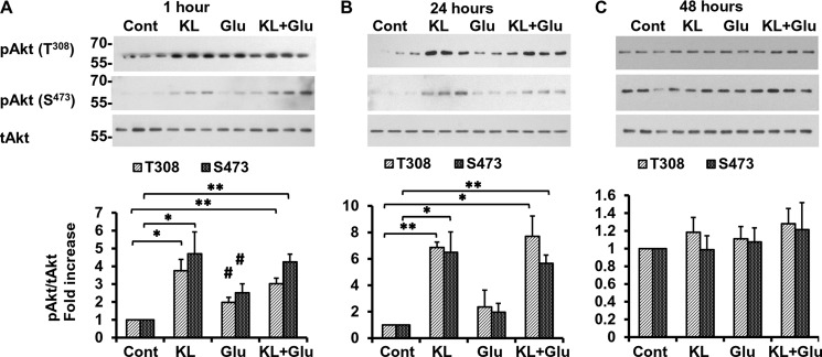
Klotho induces the phosphorylation of Akt. Rat primary hippocampal neurons were pretreated with Klotho (KL) for 4 h followed by additional treatment for 1 h (A), 24 h (B), and 48 h (C), in the absence or presence of glutamate (Glu) (3 mm). The lysates were collected for WB analysis using primary antibody recognizing the Thr308 phosphorylated site (top panels) and Ser473 phosphorylated site (bottom panels). Phosphorylated Akt proteins are normalized to total Akt (tAkt) control and presented as -fold increase normalized to untreated control. Asterisks indicate statistical significance of Klotho-treated versus Klotho-untreated samples. *, p < 0.05; **, p < 0.01; #, p < 0.05 for cells treated with glutamate only versus untreated cells by Student's t test. Error bars, S.D. The average of three independent experiments and representative blots are shown.