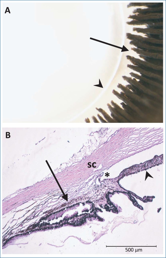
Figure 6.

(A) Gross morphology of the lens and ciliary processes viewed from the posterior side; arrowhead indicates lens equator and arrow indicates ciliary processes (B) H&E-stained section of the iridocorneal angle. Arrow indicates longitudinal ciliary muscle and arrowhead indicates posterior iris. *Angle. SC, Schlemm's canal.
