Figure 5.
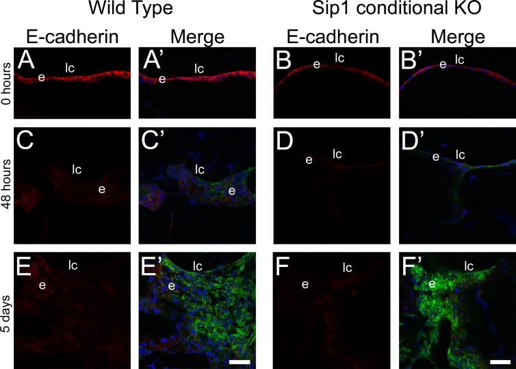
Epithelial-to-mesenchymal transition still occurs after surgery when Sip1 is not present. Confocal microscopy images of E-cadherin and α-SMA protein expression in wild-type (A, A′, C, C′, E, E′) and Sip1 knockout lenses (B, B′, D, D′, F, F′) at 0 hours (A, B), 48 hours (C, D), and 5 days (E, F) showing downregulation of E-cadherin in the 48-hour and 5-day samples in both the wild-type and Sip1 conditional knockouts. Prime panels (e.g. [A′]) show E-cadherin expression (red) merged with nuclei (DRAQ5, blue) and α-SMA expression (green). Scale bars: 35 μm. e, residual lens epithelial cells/transitioning epithelial cells.

