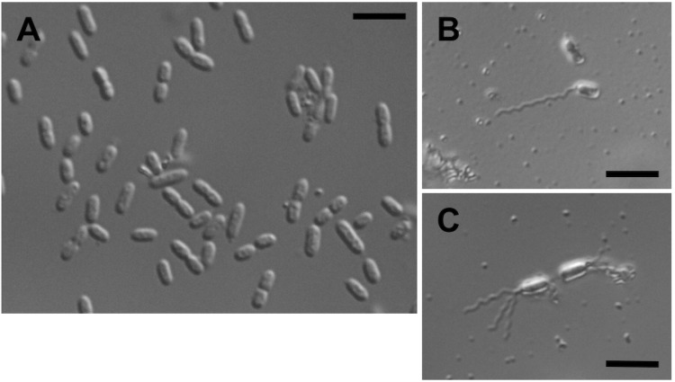
Figure 2.
Cell morphology and flagella of G. thailandicus NBRC 3257. (A) Differential interference contrast image of G. thailandicus NBRC 3257 grown on mannitol medium. Bar, 5 µm. (B and C) Microscopic images of flagella stained by the modified Ryu method. Singly (B) and multiply (C) flagellated cells were observed. Bars, 5 µm.

