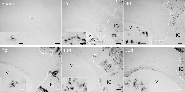
Figure 2.

Spatiotemporal expression of MHC II protein in the ischemic hemisphere. Spatiotemporal distribution of MHC II expressing cells in the ipsilateral cortex of a sham operated animal and at the indicated time points after tMCAO. The border of the infarct core is delineated by a dotted line. Scale bars: low magnification - 200 μm; higher magnification - 20 μm. Abbreviations: V, lateral ventricle; IC, infarct core.
