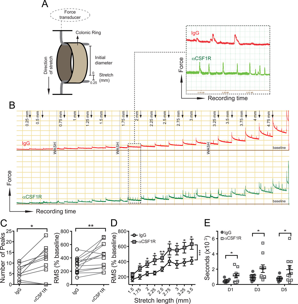
Figure 3. Depletion of MMs results in intestinal dysmotility.
(A) Illustration of ex vivo method to measure stretch-induced peristaltic contractions of colonic rings using a myograph. (B) Five hour recording of stretch-induced contractions of colonic rings during repeated application of 0.25 mm long stretching up to 5.00 mm total stretch length. 3 mm colonic rings were obtained from WT mice 2 days after i.p. injection of isotype IgG or αCSF1R mAb. (C) Number of peaks (left) or Root Mean Square (RMS) normalized to baseline in % (right) during 10 min recordings of colonic contraction at stretch distance 2.75 mm. (D) RMS normalized to baseline (%) during 10 min recordings of colonic contractions at each stretching step from 1.5 to 3.5 mm. (E) Colonic transit time measured by bead expulsion assay in WT mice 1, 3 and 5 days after i.p. injection of isotype IgG or αCSF1R mAb.

