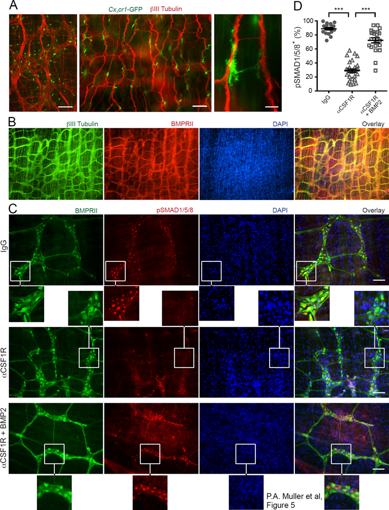
Figure 5. MMs activate BMP2 receptor on enteric neurons.
(A) Distribution of CX3CR1+ MMs in colon (left) and ileum (middle and right) muscularis from Cx3cr1-GFP+/– mice stained with anti- βIII Tubulin Ab and analyzed by IF. Scale bars – 500 nm (left), 100 nm (middle) and 10 nm (right). (B) IF analysis of LB muscularis from WT mice stained with anti-BMPRII and anti-βIII Tubulin Abs and counterstained with DAPI. Scale bars – 500 nm. (C) IF analysis of LB muscularis from WT mice 2 days after i.p. injection of isotype IgG (top), αCSF1R mAb (middle and bottom) stained with anti-pSMAD1/5/8 and anti-BMPRII Abs and counterstained with DAPI. The bottom panel shows pSMAD1/5/8 distribution in the muscularis from αCSF1R mAb injected mouse that was incubated with BMP2 as described in D. Scale bars – 100 nm. (D) Quantitative summary of the distribution of pSMAD1/5/8+BMPRII+ neurons in the muscularis from WT mice 2 days after i.p. injection of isotype IgG or αCSF1R mAb. In all cases, the muscularis was incubated for 30 min at 37°C in complete medium in the presence or absence of BMP2 (10 ng/ml) as indicated. Each data point represents % of pSMAD1/5/8+ neurons among total BMPRII+ neurons in each visual field; each column summarizes the results from three animals.

