Figure 4.

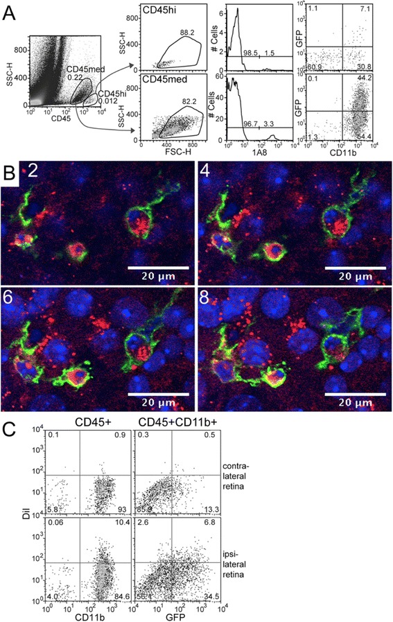
Detection of retinal ganglion cell (RGC) phagocytosis by GFP hi and GFP lo CD11b + cells in CDG retina post-ONC. (A) Depiction of CD45hi and CD45med gating, elimination of 1A8+ PMN, and selection for CD11b+ cells that are GFPlo or GFPhi. (B) Sequential confocal sections of GFPhi DC engulfing DiI-labeled RGC seven days post-ONC. DiI-labeled RGC, red; GFPhi DC, green; DAPI-stained nuclei, blue. (C) Detection and quantitation of DiI-labeled CD45medCD11b+GFPhi DC and GFPlo cells by flow cytometry. Ipsilateral retinas were labeled by injection of DiI into the superior colliculus. After seven days, mice were given an ONC; retinas were harvested six days later.
