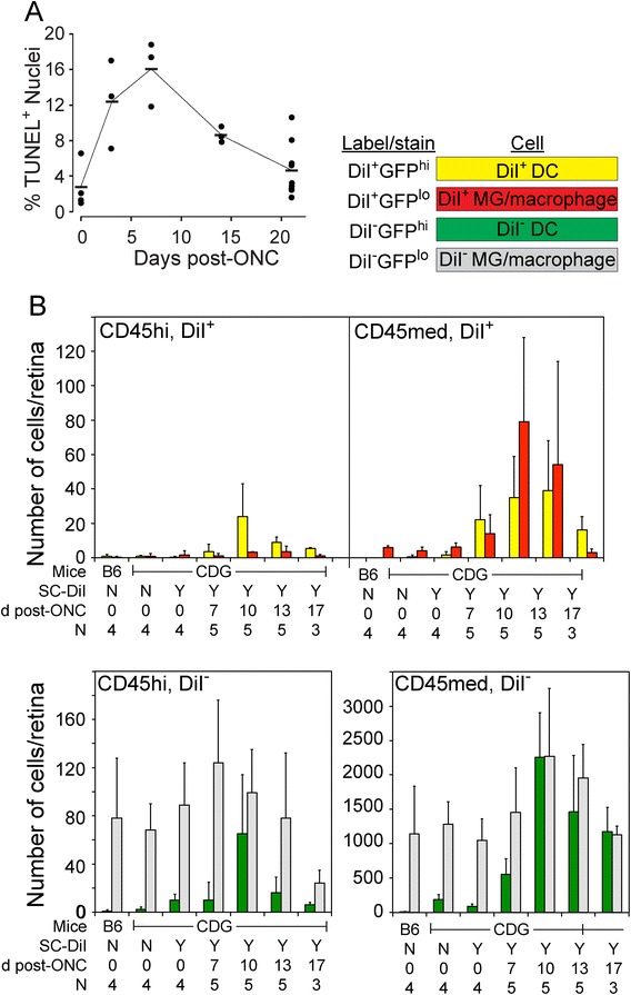
Figure 5.

Phagocytosis of retinal ganglion cells (RGC) following an optic nerve injury (ONC). (A) Time course of RGC apoptosis following an ONC. (B) Analysis of DiI uptake in GFPhi DC and GFPlo MG/macrophages following an ONC. Retinas were labeled by injection of DiI into the superior colliculus. After 7 days the mice were given an ONC; retinas were harvested 7, 10, 13, and 17 days later. CD45+ cells were examined by flow cytometry as shown in Figure 4. All cells in (B) are CD11b+.
