
Figure 7.
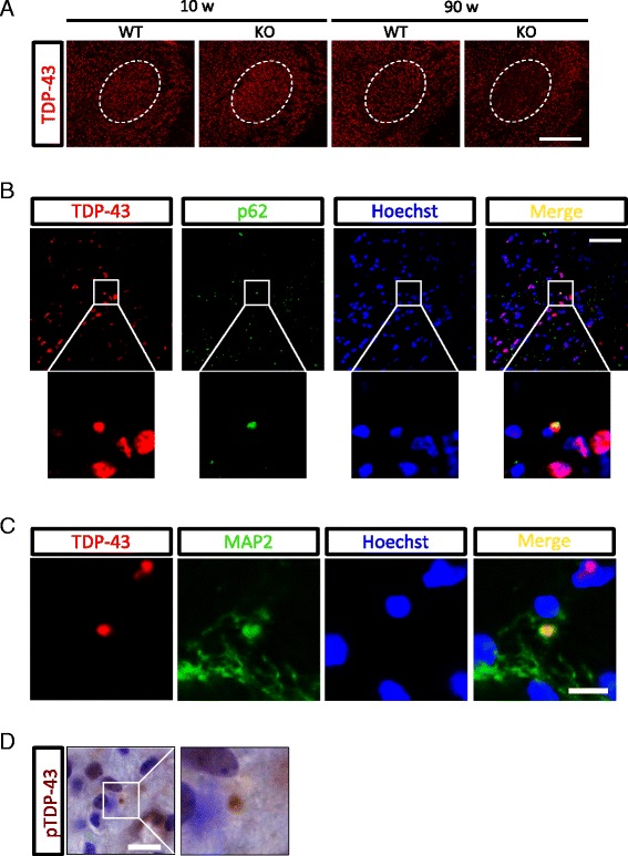
TDP-43 aggregation in the cytoplasm of neurons in the VPM/VPL of aged PGRN-deficient mice. A, TDP-43-immunostained images in the VPM/VPL of 10- and 90-week-old WT and KO mice (scale bar = 500 μm). The area enclosed by dashed line indicates the VPM/VPL. B, The triple-stained image of TDP-43 (red), p62 (green), and Hoechst33258 (blue) in the VPM/VPL of 90-week-old KO mice (scale bar = 50 μm). C, The triple-stained image of TDP-43 (red), MAP2 (green), and Hoechst33258 (blue) in the VPM/VPL of 90-week-old KO mice (scale bar = 20 μm). D, Phospho-TDP-43 (pTDP-43)-immunostained image with Nissl counterstaining in the VPM/VPL of 90-week-old KO mice (scale bar = 10 μm).