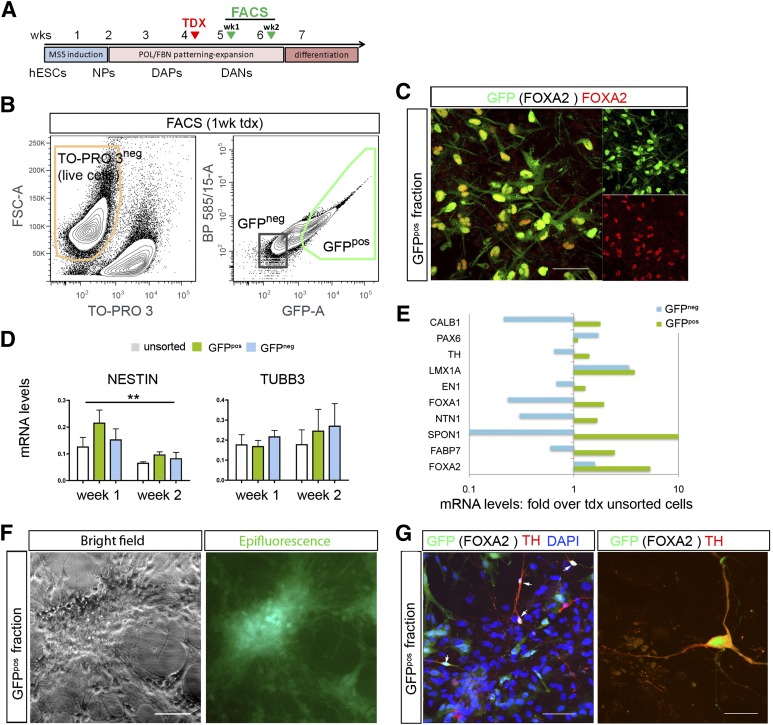
Figure 3.
Selection of human dopamine progenitors transduced with the lentiviral vector pFOXA2.GFP. (A): Schematic panel showing the protocol of human pluripotent stem cell differentiation with cells transduced at week 4 and selected by FACS 1 or 2 weeks later. (B): Representative gating strategy for isolation of viable GFPpos cells (shown is a selection of H9-derived cells, 1 week after transduction) (see supplemental online Fig. 5 for methodological details and postsort analysis). (C): Postsorting immunofluorescence (IF) confirmed a good overlap of GFP and FOXA2 (∼90%). (D): Analysis of nestin and βIII tubulin at 1 and 2 weeks in the unsorted, GFPpos, and GFPneg fractions by quantitative real-time polymerase chain reaction immediately after FACS. Shown are pooled data from 11 independent experiments. There was a significant decrease in nestin in the 2 weeks (p < .01). (E): Transcriptional profiles for floor-plate, DA, neural, and neuronal genes in the FACS cellular fractions showed enrichment for midbrain floor plate transcripts in the GFPpos fraction; data are shown here as fold change relative to unsorted cells for the corresponding transduction experiments, in a log scale for clarity. (F): Isolated GFPpos fractions could be maintained in vitro when plated at high cell densities in coculture with rat striatal astrocytes. (G): Analysis by IF of the GFPpos cells plated on astrocytes (GFP negative nuclei) showed few THpos cells that were GFPpos (arrows) at 2 weeks postsorting. Scale bars = 50 μm in (C), (F), and left panel in (G) and 20 μm for right panel in (G). Abbreviations: DANs, dopamine neurons; DAPs, dopamine progenitors; DAPI, 4[prime],6-diamidino-2-phenylindole; FACS, fluorescence-activated cell sorting; FSC, forward scatter; GFP, green fluorescent protein; hESC, human embryonic stem cell; NPs, neural progenitors; POL, polyornithine; tdx or TDX, transduction; TH, tyrosine hydroxylase; wk, week.

