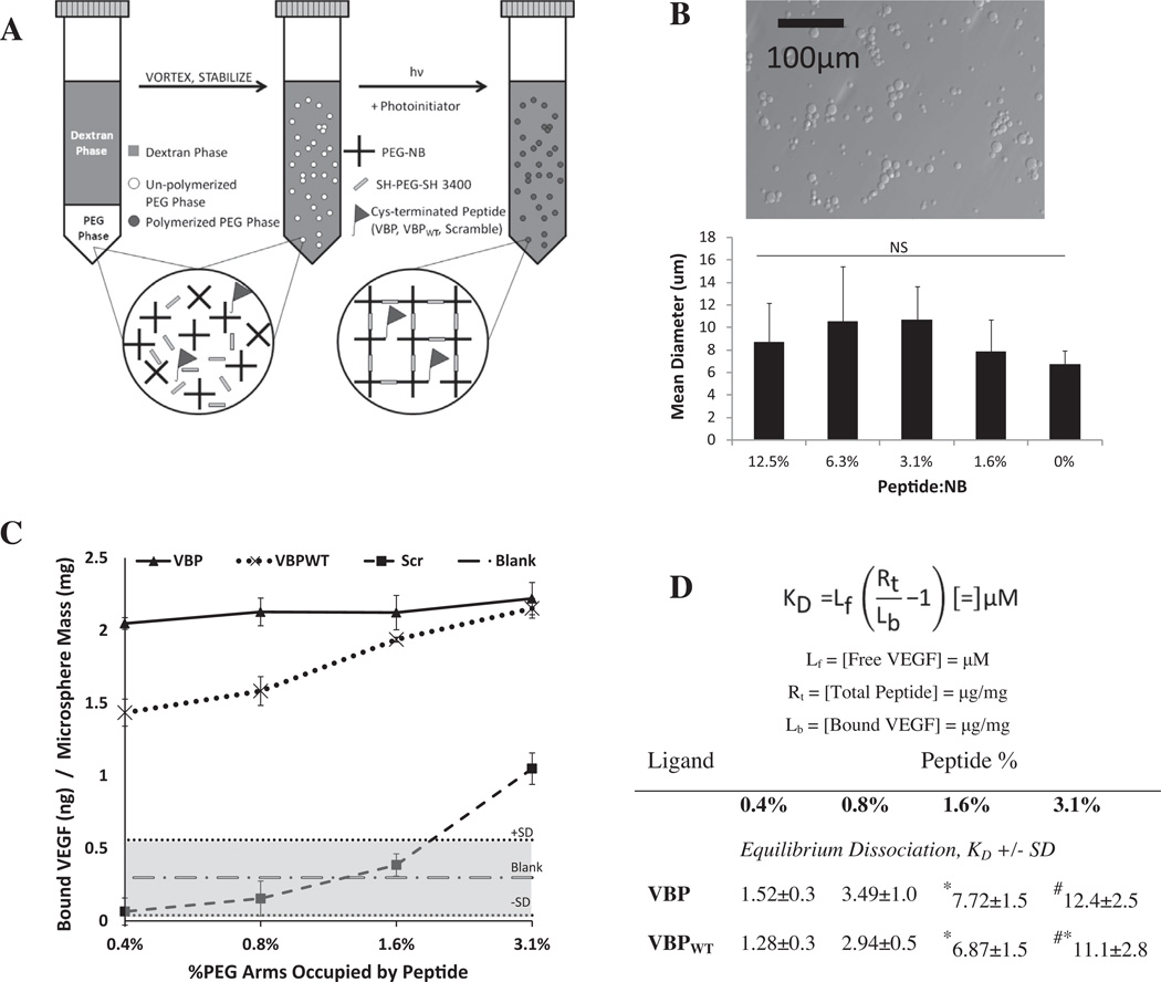
Fig. 1.
(A) Schematic of PEG-NB microsphere synthesis for covalent peptide incorporation. Shown is the emulsion procedure for synthesizing PEG microspheres using thiolene chemistry. (B) (top) Bright-field image of Blank PEG microspheres; the scale bar represents 100 µm. (bottom) Graph of mean microsphere diameter ±1 SD at a range of incorporated peptide concentrations, as determined using ImageJ software. (C, D) Comparison of peptide density and affinity, and their effect on VEGF binding at 1 mg ml−1 microspheres and 10 ng ml−1 recombinant human VEGF in 0.1% BSA in PBS loading solution. (C) Amount of VEGF bound per weight of microspheres with covalently tethered Scramble microspheres (dashed line, boxes), VBPWT microspheres (dotted line, Xs) and VBP microspheres (solid line, triangles). Sequestered VEGF for Blank microspheres is shown (alternating dashes and dots), with the gray area outlining the average ±1 SD. (D) Effective dissociation constant, KD, of microsphere binding to soluble VEGF. Results are indicated as KD ± SD, with symbols indicating a significant difference compared to 0.4% (*), 0.8% (#) and 1.6% (&) of each respective peptide at a p < 0.05.

