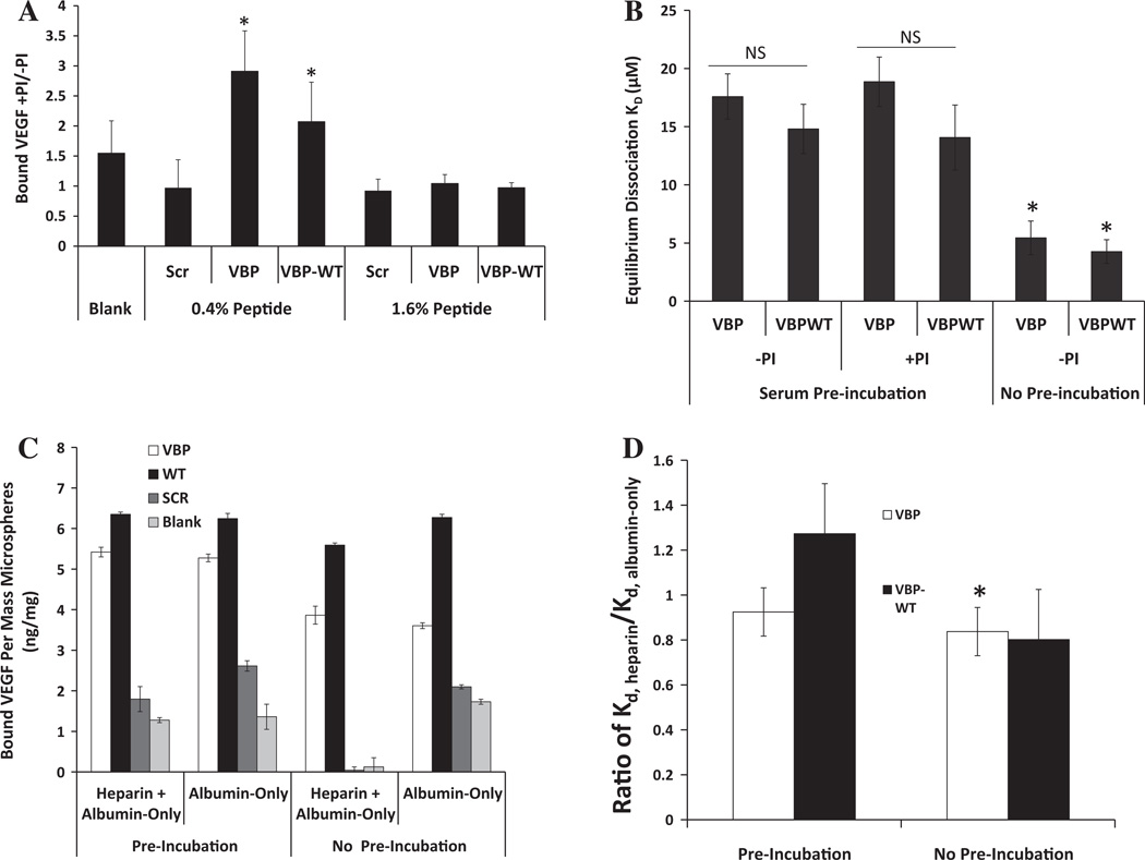
Fig. 3.
Comparison of VEGF binding with VBP and VBPWT microspheres in the presence and absence of protease inhibitor. (A) Microspheres at 1.6 and 0.4% peptide densities, as well as Blank microspheres, were incubated in 25% FBS with and without protease inhibitor cocktail (PI). Data are presented as bound VEGF (ng VEGF per mg microspheres) with protease inhibitor (+PI) divided by bound VEGF without protease inhibitor (—PI). A one-tailed Student’s t-test was performed to test if these values were significantly greater than 1, with asterisks denoting p < 0.05. (B) Comparison of effective dissociation constant for VBP and VBPWT microspheres preincubated in 25% serum with or without protease inhibitor. A two-tailed Student’s t-test was performed between VBP and VBPWT microspheres with no preincubation vs. each respective condition with serum preincubation. Asterisks denote p < 0.05. (C) Comparison between different UFH-containing solutions both in a UFH or albumin-only preincubation step or with UFH or albumin-only in the presence of VEGF. Data are presented as bound VEGF per mass of 1.6% peptide microspheres in ng mg−1 for VBP (white bars), VBPWT (black bars), Scramble (dark gray bars) or Blank (light gray bars) microspheres. No statistical significance existed between preincubation conditions, and in all cases VBP and VBPWT microspheres bound significantly more VEGF than Scramble and Blank. (D) Comparison of VEGF-binding dissociation constants of heparin-containing medium, KD,heparin, divided by the same of albumin-only medium, KD,albumin-only, for both preincubated microspheres and for no preincubation. Statistical significance is depicted as significantly less than 1 with a one-tailed Student’s t-test, with asterisks denoting p < 0.05.

