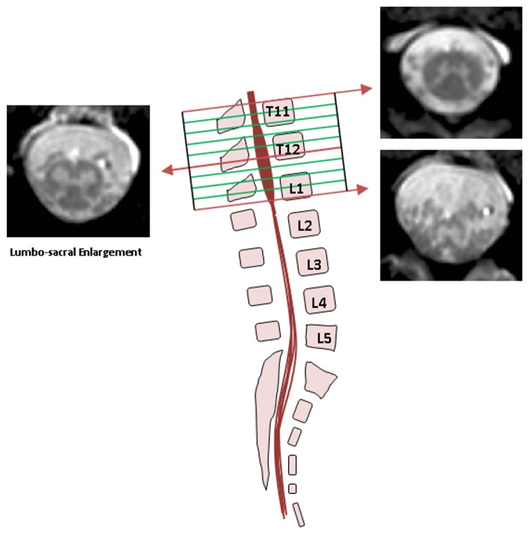
Figure 1. Imaging the lumbosacral enlargement (LSE).

The imaging volume was prescribed to cover from the superior margin of T11 to the inferior margin of the L1 vertebral bodies so that to ensure coverage of the LSE in all subjects. Example images are shown from superior, middle and inferior sections of the imaging volume.
