
Figure 1.
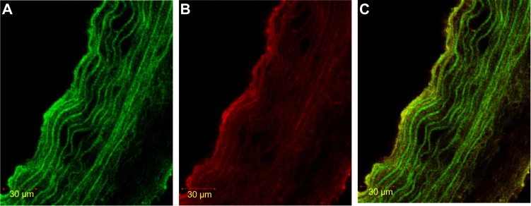
Immunofluorescence staining of HIF-1α in SMC, labeled with α-actin.
Notes: (A) α-actin with FITC. The color green shows the expression of α-actin (marker SMC). (B) HIF-1α rhodamine. Red shows HIF-1α expression. (C) Double stained α-actin FITC – HIF-1α. Yellow shows double staining of HIF-1α expression in SMC using. rhodamine.
Abbreviations: FITC, fluorescein isothiocyanate; HIF-1α, hypoxia-inducible factor-1α; SMC, smooth muscle cell.