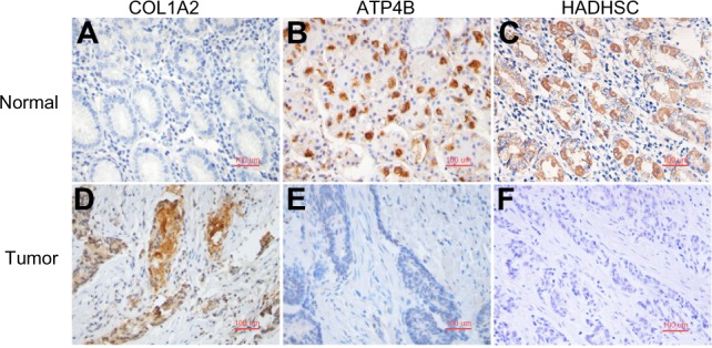
Figure 2.

Validation of the feature genes using IHC staining. (A) and (B): positive staining of COL1A2 appeared in cancer but not in NT. COL1A2 was highly expressed in 17 GC samples with the positive rate of 77.3% (17/22). (C) and (D): negative staining of ATP4B appeared more often in cancer but positive in NT. ATP4B was highly expressed in 20 normal samples with the positive rate of 83.4% (20/24). (E) and (F). positive staining of HADHSC appeared in normal but not in cancer tissue. HADHSC showed 24 normal samples with high expression of 92.3% positivity (24/26).
